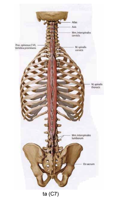
The skeleton is divided into the spine, chest, upper/lower extremities, and skull. Spine (Columna vertebralis) Consists of a total of 33-34 vertebrae.
Functions of the spine: stability, support, cushioning, shock absorption, and protection of the spinal cord.
7 cervical vertebrae (vertebra cervicalis)
12 thoracic vertebrae (vertebra thoracica)
5 lumbar vertebrae (vertebra lumbalis)
5 sacral vertebrae (vertebra sacralis)
4-5 coccygeal vertebrae (vertebra coccygis)
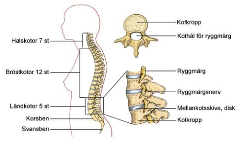
+ intervertebral discs (discus intervertebralis)
The natural S-shape of the spine is caused by:
- a cervical lordosis
- a thoracic kyphosis
- a lumbar dose
- a tailfish
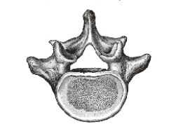
The vertebrae
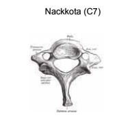
A vertebra is made up of a vertebral body, vertebral arch, and processes and joints. The vertebral body is well developed in the lumbar spine, less developed in the cervical spine. The vertebral arch is well developed in the cervical spine, less developed in the lumbar spine. The most developed processes are the transverse processes (proc. transversus) and spinous processes (proc. spinosus). Many processes (
) are in contact with the vertebrae or ribs above or below, thus forming joints.
The chest (Thorax)
Structure
- Twelve pairs of ribs (costae), ten of which connect to the sternum. The two lowest ribs have no anterior contact with the sternum.
- The top seven ribs connect directly via the costal cartilage, while ribs 8-10 connect indirectly to the sternum via the costal arch.
- The sternum consists of three bones: the manubrium, the sternum (corpus sterni), and the xiphoid process (proc. xiphoideus).
- Twelve thoracic vertebrae with joints toward the ribs dorsally.
Function
- Protect internal organs.
- Create space for lungs and breathing
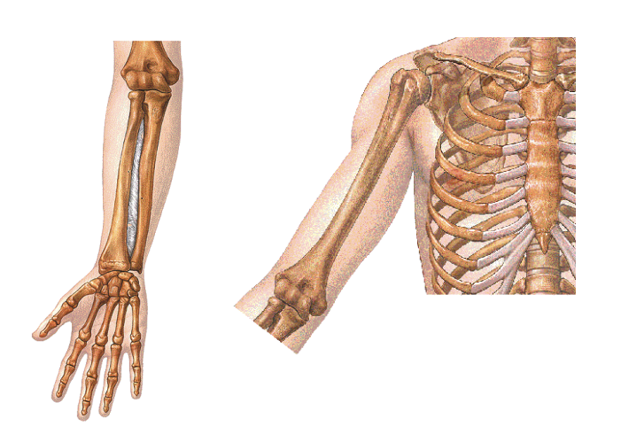
Skeleton of the upper extremities
The shoulder girdle is made up of the shoulder blade (scapula), which connects to the collarbone (clavicle). The collarbone also connects to the manubrium sterni. The scapula has a joint socket that forms the attachment point for the upper arm bone (humerus). The humerus leads distally to the radius and ulna. Both the radius and ulna lead to the carpal bones. These lead to the metacarpal bones, which lead to the phalanges of the fingers.
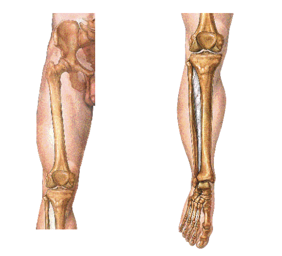
Skeleton of the lower extremities
Just like the upper extremities, the lower extremities are also made up of a proximal bone, two distal bones, a proximal ball joint, and a distal hinge joint. The femoral head (caput femoris) articulates with the acetabulum in the pelvis. Femoral neck (collum femoris) Connects the femoral head to the body of the femur (corpus femoris). Distally, the femur forms the upper part of the knee joint (articulatio genus), while the lower part is formed by the tibia. In front of the knee joint is the kneecap (patella). Distally, the tibia together with the fibula form the upper part of the ankle joint. The foot is made up of the tarsal bones, the metatarsal bones, and the toes (phalanges).
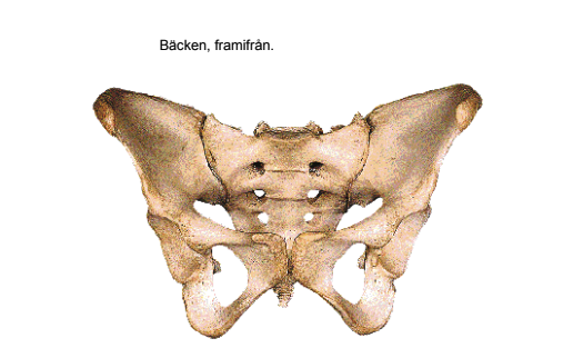
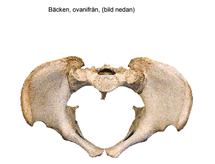
The pelvis
The pelvis consists of the coccyx, two hip bones (os coxae) that connect dorsally to the sacrum and anteriorly to a cartilaginous joint (symphysis). Each hip bone consists of three fused bone parts: the ilium, the ischium, and the pubis. Together, these form the acetabulum, which connects to the femur. Women have a larger pelvic girdle than men. During pregnancy, the joints in the pelvis loosen up (symphysis pubis diastasis), which facilitates the delivery of a child.
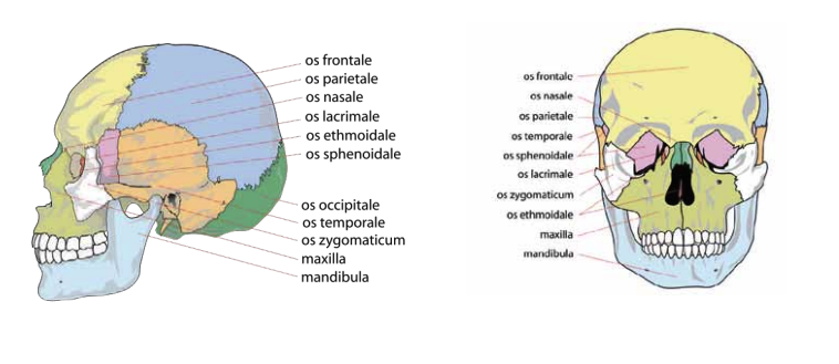
The skeleton of the head
Consists of 29 partially fused flat bones. The main functions of the skull bones are to protect the brain and sensory organs. The skeleton of the head can be divided into the cranium and the facial skeleton.
The largest bones in the cerebral skull:
Occipital bone – os occipitale
Sphenoid bone – os sphenoidale
Ethmoid bone – os ethmoidale
Temporal bone – os temporale
Parietal bone – os parietale
Frontal bone – os frontale
Facial skull:
Nasal bone – os nasale
Cheekbone – os zygomaticum
Upper jawbone – os maxillaris (maxilla)
Lower jawbone – os mandibularis, (mandibula)