Muscle Anatomy and Physiology
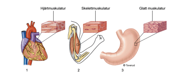
There are three types of muscle tissue in the body: skeletal, cardiac, and smooth muscle. The muscles involved in RAPP are composed of skeletal muscle cells and account for approximately 45% of body weight. Skeletal muscles are not only used by RAPP but also for functions such as breathing and swallowing. Each individual skeletal muscle is attached to the skeleton at at least two points. In most cases, the muscle attaches to different sides of a joint, so a contraction of the muscle causes movement in the joint.
Our skeletal muscle system consists of approximately 300–400 striated muscles. It is the skeletal muscles that enable us to move. Most of these are paired, and almost everywhere on the body, the muscles are arranged in multiple layers. Muscles account for about 40% of body weight, but regular muscle training increases muscle tissue and thus weight, while reduced muscle training has the exact opposite effect.
The naming of muscles is rather unsystematic. Some muscles are named after their shape or size, others after their location, while still others are named after the number of “heads” they have. Our skeletal muscles cannot perform any work to produce movement on their own; they can only contract. To produce movement, muscles must work in conjunction with the bones of the skeleton. A muscle and a bone together form a lever, with the moving bone acting as the fulcrum. Muscle tissue is never directly attached to the skeleton but is always connected via a tendon made of connective tissue. In addition to the skeleton, a muscle can also be attached to: skin, ligaments, joint capsules, etc.
A muscle never works alone but always interacts with several other muscle groups. This brings us to a new classification of muscles: agonists, antagonists, and synergists. Let’s take a common movement—bending the arm—as an example. When the biceps muscle on the front of the upper arm bends the arm at the elbow joint, the extensor muscle (m. triceps brachii) on the back of the upper arm does not relax suddenly, but instead develops a gradually decreasing pulling force in the opposite direction. This gives the movement a smooth, gliding motion. The biceps brachii muscle, which performs the movement, is called the agonist, while the triceps brachii muscle, which opposes the movement, acts as the antagonist. Other upper arm muscles that contribute to the flexion and control of the forearm and assist the biceps brachii are called synergists.
The Structure of Skeletal Muscles
Skeletal muscle cells consist of several cells that have fused together. Each cell therefore contains multiple nuclei.
After birth, no new skeletal muscle cells are formed. Instead, as we grow, the muscle cells increase in size. Some muscle cells can grow up to 30 centimeters long. Skeletal muscle cells contain protein filaments arranged in a very regular pattern, which gives the muscle tissue a striated appearance when viewed under a microscope.
There are two types of protein filaments: actin and myosin. These filaments can shift relative to one another, so that they are sometimes completely interlocked and sometimes more widely spaced. When the filaments are interlocked, the muscle cell shortens. This is what happens when the muscle contracts or tenses.
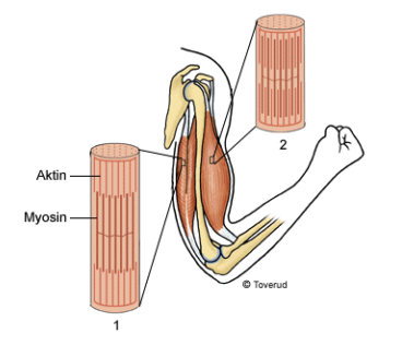
Skeletal muscles contain thin filaments of two different types of protein: actin and myosin. These protein filaments can slide past one another. When the muscle is relaxed, the protein filaments are separated from one another (1). When the muscle contracts, the actin and myosin filaments slide past one another (2).
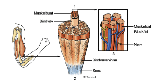
Muscle cells are arranged in parallel bundles held together by connective tissue (1). Several bundles together form a muscle. A slightly thicker layer of connective tissue surrounds the entire muscle and extends into the muscle’s tendons (2). The muscles receive oxygen and nutrients from blood vessels that enter the muscle. The blood vessels follow the connective tissue between the muscle cells. Nerves also run through the connective tissue, transmitting information to and from the muscle (3)
Type I and Type II muscle fibers/cells
We now know that there are two different types of muscle fibers: slow-twitch (Type I) and fast-twitch (Type II). Slow-twitch fibers obtain their energy from oxygen in the blood. Fast-twitch fibers derive most of their energy from glucose stored in the muscle.
Type I fiber training
Based on research conducted at relatively low intensity and over a long period of time, it has been found that type I fibers respond with the following changes:
- The capillary network in the muscle and its ability to supply oxygen to the muscle increase, particularly around type I fibers. (aerobic)
- The number of mitochondria in muscle cells increases, and their function is to release energy (ATP).
- The number of repetitions at a submaximal level increases.
Type II fiber training
High-intensity training primarily affects type II fibers. The main changes are as follows:
- The cross-sectional area of the cells increases, thereby enhancing their ability to generate more power.
- The cells' ability to function without oxygen (anaerobically) increases.
- Neuro-muscular coordination improves. More motor units are activated (technique).
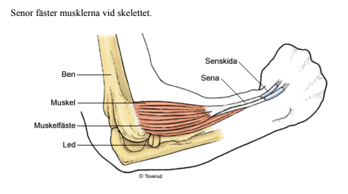
Senor
Tendons attach muscles to the skeleton
The connective tissue sheath that surrounds each skeletal muscle transitions at the muscle’s end into a tendon that attaches to the bone. Tendons consist of dense connective tissue. The shortest tendons are only a few millimeters long, and the longest are about 30 centimeters.
At the point where it attaches to the skeleton, the tendon’s connective tissue is interwoven with the bone tissue. This creates a very strong attachment. The force generated in the muscle as it contracts is transferred to the tendon and on to the skeletal structures, causing them to move.
Tendons are easily susceptible to wear and tear. In many places, especially where the tendon glides over a hard bony edge, there are tendon sheaths or bursae that protect the tendons. The tendons of the hand and foot are particularly vulnerable and therefore run through tendon sheaths. Bursae may be connected to joint cavities. Both tendon sheaths and bursae contain fluid that reduces friction. Tendons can also be held in place by ligaments.