The muscles of the leg
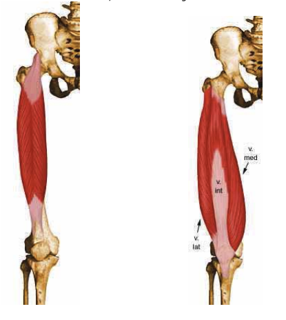
M. Quadriceps femoris (four-headed thigh muscle)
Source:
M. Rectus femoris Origin (O): Anterior inferior iliac spine. Insertion:
(I) Patella and tibial tuberosity.
M. vastus lateralis O: Greater trochanter, lateral linea aspera (lateral/posterior femur)
F: Lateral aspect of the patella, tendon of the rectus femoris, tibial tuberosity (Tt)
Vastus medialis muscle U: Medial/posterior aspect of the femur.
F: Medial aspect of the patella, medial aspect of the tendon of the rectus femoris
Vastus intermedius muscle U: Anterior aspect of the femur. F: Lower part of the patella. The vastus lateralis, intermedius, and medialis merge to form the patellar ligament, which in turn attaches to the tibial tuberosity (anterior aspect of the proximal tibia).
Function: Knee extension; the rectus femoris also performs hip flexion.
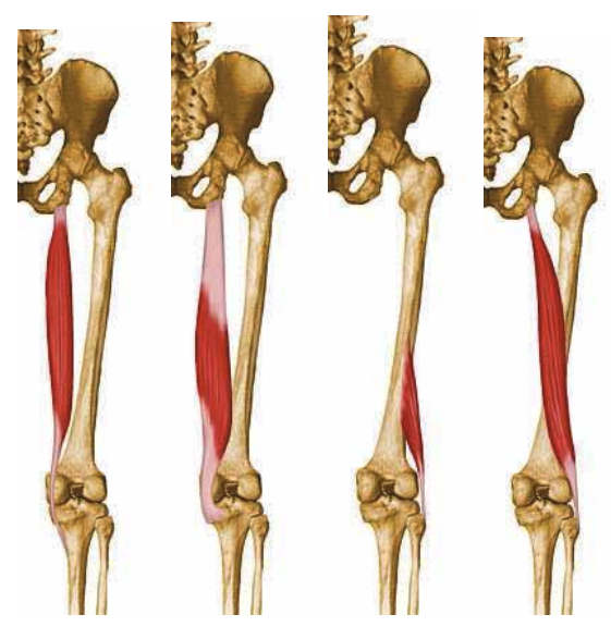
Hamstrings (back of the thigh)
M. Biceps femoris. U: ischial tuberosity, posterior part of the femur; F: head of the fibula
M. Semitendinosus. U: ischial tuberosity; F: pes anserinus (medial aspect of the proximal tibia)
M. Semimembranosus. U: ischial tuberosity; F: medial aspect of the tibia, partially covered by the semitendinosus.
Function: flexion of the knee joint and extension of the hip (all 3), as well as external rotation of the knee (biceps) and internal rotation (semimembranosus and semitendinosus) of the knee and hip joints, and adduction of the hip joint.
Images from left: Semitendinosus, Semimembranosus, short head of the biceps, and long head of the biceps.
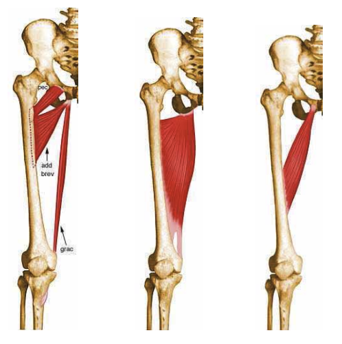
Adductors (muscles that bring the leg inward)
M. Gracilis Origin: os pubis (medial to the adductor magnus) Insertion: pes anserinus. Adduction, internal rotation of the hip joint, flexion, internal rotation of the knee joint
M. Pectineus Origin: os pubis Insertion: inferior to the lesser trochanter. Adduction, flexion, external rotation of the hip joint
M. Adductor longus Origin: os pubis Insertion: linea aspera (medial/posterior femur). Adduction and flexion of the hip joint
M. Adductor brevis Origin: os pubis Insertion: linea aspera (medial/posterior femur). Hip adduction and external rotation
M. Adductor magnus U: Tuber ischiadicum (os pubis, lateral to the other adductors),
F: medial femoral condyle, linea aspera. Hip adduction
Figure 1: Adductor brevis, pectineus, and gracilis. Figure 2: Adductor magnus. Figure 3: Adductor longus.
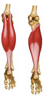
M. Triceps surae (three-headed calf muscle)
M. Gastrocnemius Origin: Proximal to the medial and lateral femoral condyles (medial and lateral heads, respectively)
M. Soleus Origin: Posterior to the proximal parts of the tibia and fibula.
They share an attachment to the calcaneal tubercle (heel bone) via the Achilles tendon.
Function: plantar flexion and supination of the foot; flexion of the knee (gastrocnemius only)
Lateral and medial gastrocnemius on the left, soleus on the right.
Extensor muscles of the lower leg
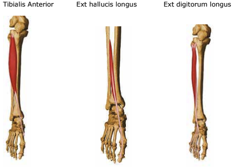
M. Tibialis Anterior
Origin: Proximally , on the anterior/lateral aspect of the tibia, membrana interossea
Insertion: Medial aspect of the medial cuneiform bone and the first metatarsal bone.
Function: Primarily dorsiflexion of the ankle joint, but also supination, since the origin is lateral and the insertion on the foot is medial.
M extensor digitorum longus
Origin: Lateral tibial condyle, interosseous membrane, fibula
Insertion: Dorsal aponeuroses (tendon sheaths) ofthe second through fifth toes
Function: Dorsiflexion and pronation of the ankle joint
M. extensor hallucis longus
Origin: Membrana interossea
Insertion: Distal phalanx of the big toe
Function: Dorsiflexion of the foot, extension of the big toe.